
Ofbyld:Computed tomography of human brain (1).png
Gjin hegere resolúsje beskikber.
Computed_tomography_of_human_brain_(1).png (512 × 512 pixels, bestânsgrutte: 79 KB, MIME-type: image/png)
| Beskriuwing |
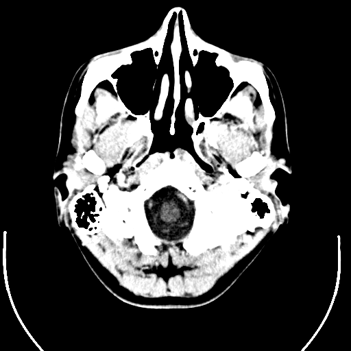
English: Computer tomography of human brain, from base of the skull to top. Taken with intravenous contrast medium.
It was taken Mars 23, 2007 on the uploader, after a 20 minute episode of homonymous hemianopsia with loss of the left visual field, but nothing strange was found. Three episodes of scotoma occurred in the following years, whereof the last one was scintillating (depiction). Otherwise, there were no further neurological symptoms.
Türkçe: Geçirdiği bir kaza neticesinde homonim hemianopsi vakası oluşan bir hastanın beyninin bilgisayarlı tomografisi. Tomografi neticesinde bir anomaliye rastlanmamıştır. |
| Datum | Uploaded January 17, 2008 |
| Boarne | Radiology, Uppsala University Hospital. Uploaded by Mikael Häggström. |
| Auteur | Department of Radiology, Uppsala University Hospital. Uploaded by Mikael Häggström. |
| Tastimming (Reusing this file) |
Compound images
-

-
 Inverted


Scrollable stack
For larger version, see Category:Computed tomography images of Mikael Häggström's brain. To move through the images, hover over the image and use scroll wheel, drag the mouse, or click the < or the > above each stack. This functionality should activate when the page is fully loaded, which may take some time.

.png)
.png)
.png)
.png)
.png)
.png)
.png)
.png)
.png)
.png)
.png)
.png)
.png)
.png)
.png)
.png)
.png)
.png)
.png)
.png)
.png)
.png)
.png)
.png)
.png)
.png)
.png)
.png)
.png)
.png)
.png)
.png)
.png)
.png)
.png)
Case with multiplanar reconstruction
-
 Brain, case 1: Multiplanar, but no intravenous contrast.
Individual images
Licencing
| This file is made available under the Creative Commons CC0 1.0 Universal Public Domain Dedication. | |
| The person who associated a work with this deed has dedicated the work to the public domain by waiving all of their rights to the work worldwide under copyright law, including all related and neighboring rights, to the extent allowed by law. You can copy, modify, distribute and perform the work, even for commercial purposes, all without asking permission.
|
DICOM format
Triemskiednis
Klik op in datum/tiid om it bestân te besjen sa't it op dat stuit wie.
| Datum/Tiid | Miniatuer | ôfmjittings | Meidogger | Opmerking | |
|---|---|---|---|---|---|
| lêste | 1 feb 2008, 14.24 | | 512 × 512 (79 KB) | Mikael Häggström | {{34 computer tomography images}} |
Bestânsgebrûk
De neikommende side brûkt dit bestân:
Globaal bestânsgebrûk
De neikommende oare wiki's brûke dit bestân:
- Gebrûk op nl.wikipedia.org
.png)